Anatomy and Physiology of Larynx , Action of Laryngeal muscles , Dr Bhanu prakash
Hellow guys, Welcome to my website, and you are watching Anatomy and Physiology of Larynx , Action of Laryngeal muscles , Dr Bhanu prakash. and this vIdeo is uploaded by Dr.G Bhanu Prakash Animated Medical Videos at 2017-07-29T00:06:39-07:00. We are pramote this video only for entertainment and educational perpose only. So, I hop you like our website.
Info About This Video
| Name |
Anatomy and Physiology of Larynx , Action of Laryngeal muscles , Dr Bhanu prakash |
| Video Uploader |
Video From Dr.G Bhanu Prakash Animated Medical Videos |
| Upload Date |
This Video Uploaded At 29-07-2017 07:06:39 |
| Video Discription |
📌 𝐅𝐨𝐥𝐥𝐨𝐰 𝐨𝐧 𝐈𝐧𝐬𝐭𝐚𝐠𝐫𝐚𝐦:- https://www.instagram.com/drgbhanuprakash
📌𝗝𝗼𝗶𝗻 𝗢𝘂𝗿 𝗧𝗲𝗹𝗲𝗴𝗿𝗮𝗺 𝗖𝗵𝗮𝗻𝗻𝗲𝗹 𝗛𝗲𝗿𝗲:- https://t.me/bhanuprakashdr
📌𝗦𝘂𝗯𝘀𝗰𝗿𝗶𝗯𝗲 𝗧𝗼 𝗠𝘆 𝗠𝗮𝗶𝗹𝗶𝗻𝗴 𝗟𝗶𝘀𝘁:- https://linktr.ee/DrGBhanuprakash
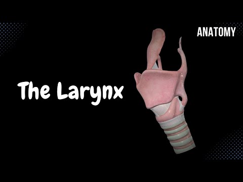
Upper Airway: Larynx
The larynx is organized into 3 major regions
1) Vestibule: between the entrance to the larynx and the vestibular folds (i.e. “false vocal cords”).
The vestibular folds contain the vestibular ligaments which are the thickened inferior edges of the quadrangular membrane.
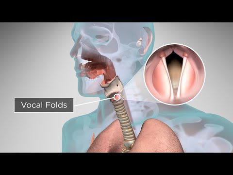
2) Ventricles: The portion between the false vocal cords (superiorly) and the true vocal folds (inferiorly).
The vocal folds contain the vocal ligaments which are thickenings of the superior edge of the conus elasticus.
Vibration of the adducted vocal ligaments with expiration produces sound (see section on muscles below for more on movement of the vocal ligaments).
3) Infraglottic cavity: Portion of the larynx inferior to the vocal folds. It communicates distally with the lumen of the trachea.
The larynx is composed of a cartilaginous skeleton
Thyroid cartilage: Composed of 2 laminae and 2 pairs of cornua.
Laminae: The plates which form the majority of the thyroid cartilage. The indentation in the midline is known as the superior thyroid notch. The inferior pharyngeal constrictor, sternothyroid and thyrohyoid muscles all attach to the laminae.
Cornua: Fingerlike projections that extend superiorly and inferiorly from the laminae.
Thyrohyoid membrane: Connective tissue membrane which connects the thyroid cartilage to the hyoid bone.
Cricoid cartilage: Composed of a narrow anterior arch and a broad posterior lamina.
Arch: Connects to the thyroid cartilage superiorly via the median cricothyroid ligament and to the first tracheal ring inferiorly via the cricotracheal ligament.
Lamina: Articulates superiorly with the arytenoid cartilages and the inferior cornua of the thyroid cartilage.
Clinical Correlate: In the event of an emergency if an airway can not be established by endotracheal intubation a cricothyroidotomy can be done where the cricothyroid membrane is incised.
Arytenoid cartilages: Pyramidal cartilages that articulate at their base with the lamina of the cricoid cartilage.
Muscular process: Extends laterally and provides attachment points for muscles.
Vocal process: Extends anteriorly and is the attachment point for the vocal ligaments.
Muscles of the larynx
Cricothyroid: Tilts the thyroid cartilage forward.
Action: Tenses and adducts the vocal ligaments.
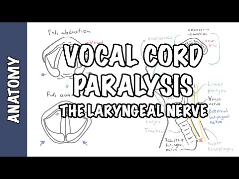
Posterior cricoarytenoid: Laterally rotates the arytenoid cartilage.
Action: Abducts the vocal ligaments.
Note: These muscles are the only abductors of the vocal ligaments and are thus extremely important to maintaining an open airway!
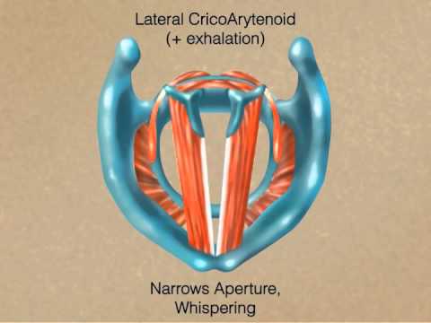
Lateral cricoarytenoid: Medially rotates the arytenoid cartilage.
Action: Adducts the vocal ligaments.
Arytenoideus: Composed of transverse and oblique parts.
Action: Adducts the vocal ligaments.
Thyroarytenoideus: From the thyroid laminae to the arytenoid cartilage.
Action: Adducts the vocal ligaments.
Thyroepiglotticus: From the thyroid laminae to the lateral aspect of the epiglottis.
Action: Holds epiglottis closed during swallowing to prevent entrance of food or liquid into the larynx during swallowing.
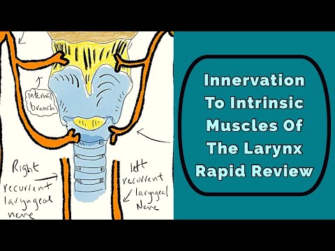
Nerves of the larynx
Superior laryngeal nerve: Divides into internal and external laryngeal nerves.
Internal laryngeal nerve: Enters thyrohyoid membrane with superior laryngeal artery. Provides sensory innervation to mucosa superior to vocal folds.
External laryngeal nerve: Travels with superior thyroid artery and provides motor innervation to the cricothyroid muscle.
Recurrent laryngeal nerves:
- Right: Loops under subclavian artery
- Left: Loops under arch of aorta
Both ascend posterior to the esophagus and enter the larynx at the level of the cricothyroid articulation.
Motor innervation to ALL muscles of the larynx (except the cricothyroids, as noted above) and provides sensory innervation to the mucosa of the larynx inferior to the vocal folds.
Arteries of the larynx
Superior laryngeal artery: A branch of the superior thyroid artery.
Travels with the internal laryngeal nerve.
Inferior laryngeal artery: A branch of the inferior thyroid artery.
Travels with the recurrent laryngeal nerve.
#larynxanatomy #larynxphysiology #anatomyoflarynx #physiologyoflarynx #larynx
#drgbhanuprakash #animatedmedicalvideos #bhanuprakashanatomylectures #usmlevideos #usmleanatomyvideos #usmlestep1videos #movementoflarynx #physiologyoflarynx #functionsoflarynx #laryngealmusclemovements #vocalcords #vocalcordmovements |
| Category |
Education |
| Tags |
usmle step 1 | larynx | physiology of laynnx | anatomy of larynx | movements of larynx | larynx animation | Anatomy and Physiology of Larynx | Action of Laryngeal muscles | laryngeal muscle action | muscles of the larynx | muscles of the larynx animation | intrinsic muscles of the larynx | extrinsic muscles of larynx | intrinsic muscles of larynx | laryngeal muscles anatomy | laryngeal muscles and their function | usmle | mbbs | larynx muscles | larynx anatomy | muscles of larynx | laryngeal muscles |
More Videos